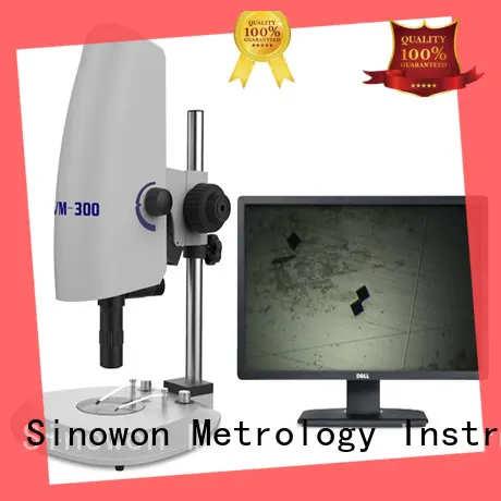
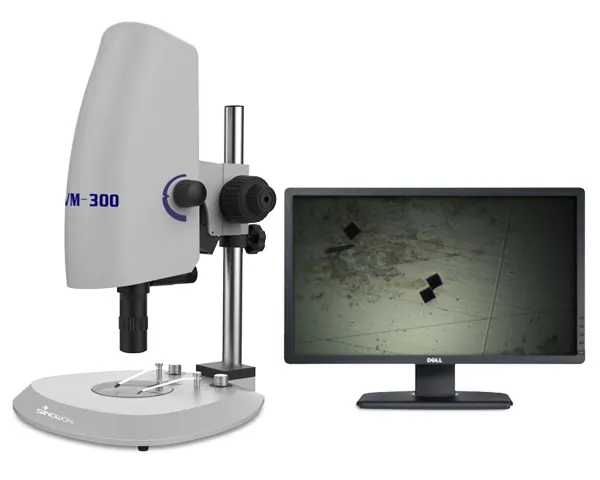
Sinowon sturdy digital optical microscope for steel products
- Sinowon provides strong guarantee for multiple aspects such as product storage, packaging and logistics. Professional customer service staff will solve various problems for customers. The product can be exchanged at any time once it is confirmed to have quality problems.
-
v s
1. Sinowon digital microscope camera is manufactured under a complete and scientific quality management system.
2. The core component of digital microscope camera is excellent, primarily shows in digital optical microscope .
3. All defects are removed from the products during the quality inspection process.
4. Due to these features, it is extensively used in varied applications.
Application:
Video Microscope is widely used in observing translucent or opaque material in electronics, metallurgy, chemical and instrumentation industry such as metal ceramic, integrated block, printed circuit boards, liquid crystal plate, film, fiber, coating and other non-metallic materials. Trinocular Video Microscope is also suitable for medicine, agriculture, forestry, public security, schools, scientific research departments for observation and analysis.
Product Feature
◆ Integral design, exquisite, fashion, generous;
◆ With high definition 0.7~4.5X horizontal zoom lens, it is more convenient and fast to switch objective magnification, and it can enlarge with high magnification with optional APO 5X/10X lens;
◆ With precision coarse and fine lifting system, ensure perfect focus and clear image;
◆ With adjustable LED bottom and surface illumination, when equipped with APO lens, it can used contour illumination and control brightness independently;
◆ Built-in FULL HD sensor and VGA integrated camera, can directly connect with the monitor to realize video image and can also use SD card to take picture or record video, which integrate video observation and image storage;
◆ Simple external interface: 12V power supply input and USB/VGA video output, SD card hole to make installation simpler.
Specification
| Product Name | Coaxial Illumination Video Microscope |
| Model | VM-300 |
| Code# | 451-430 |
| Electronic Magnification | 16X~206X (16:9 21”Monitor) |
| Optical Objective | 0.7X~4.5X horizontal zoom lens; Zoom ratio 6.5:1; 1X C-mount Camera Adapter;10X APO Lens: WD:35mm |
| Camera Parameter | 2 million pixel (1920*1080);Image Size:1/2.86”;Frames Per Second:30fps |
| Camera Function | Photograph and Video Record Cross Line VGA/USB Output, SD Card Storage OSD: Comprehensive digital UI design, Wireless USB 2.0 Mouse Operation White Balance; Brightness Control; Digital Noise Reduction Picture Frozen,10X Digital Magnification |
| Measuring Function | Optional PC System and measuring software can measure image in the field of view. |
| Illumination | Bottom: Adjustable LED Illumination;Surface: Adjustable LED Illumination |
| Microscope Stand | Z-axis Travel: 150m mCoarse/Fine Lifting System |
| Electrical Parameters | AC90~240V;50~60HZ |
| Dimension | 290*280*400 mm |
| Gross/Net Weight | 7.0/4.7Kg |
Standard Delivery:
| Product Name | Specification |
| Microscope Main Body | VM-300 |
| Power Adapter | DC12V |
| Mouse | Wireless |
| SD Card | 8G |
| Operation Manual/Warranty Card/Certificate/Packing list | VM-300 |
Camera Parameter:
| Image | Image Sensor | 1920*1080 FULL HD Sensor 1/2.86” |
| Effective Pixel | 1920*1080 | |
| Pixel Dimension | 2.75*2.75um | |
| Frame Rate | 30 | |
| Definition | FULL HD | |
| Function | White Balance | Auto, Manual |
| Digital Magnification | Support 10X zoomed in and out | |
| Brightness Control | Auto, Manual | |
| Color | R/G/B Adjustable | |
| Frozen | Support | |
| OSD | Comprehensive digital UI design, Mouse Operation | |
| Enhance Edge Definition | Support | |
| Photograph and Video Record | Support | |
| Cross Cursor | Support, multiple color optional, coarse/fine adjustable | |
| Horizontal and Vertical Line | Support, multiple color, four horizontal and vertical line, position movable, coarse line adjustable | |
| Image Comparison | Support | |
| Wide Dynamic | Support | |
| Digital Noise Reduction | Support | |
| Interface | VGA Interface | Standard VGA output |
| Storage Interface | SD Card Slot | |
| USB Interface | Standard USB2.0 Interface, can connect to mouse and PC System |
Field of View:
Auxiliary objective | Field of view (0.5X Camera Adapter) | Remark | |||||||||||||||||||
| 0.7X | 1X | 2X | 3X | 4X | 4.5X | ||||||||||||||||
| Length | Width | Diagonal | Length | Width | Diagonal | Length | Width | Diagonal | Length | Width | Diagonal | Length | Width | Diagonal | Length | Width | Diagonal | ||||
0.5X WD:108mm | Magnification 0.18X-1.12X | Optional | |||||||||||||||||||
| 25.29 | 22.86 | 34.09 | 17.70 | 16.00 | 23.86 | 8.85 | 8.00 | 11.93 | 5.90 | 5.33 | 7.95 | 4.43 | 4.00 | 5.97 | 3.93 | 3.56 | 5.30 | ||||
1.0X WD:91mm | Magnification 0.35X-2.25X | Standard | |||||||||||||||||||
| 12.64 | 11.43 | 17.04 | 8.85 | 8.00 | 11.93 | 4.43 | 4.00 | 5.97 | 2.95 | 2.67 | 3.98 | 2.21 | 2.00 | 2.98 | 1.97 | 1.78 | 2.65 | ||||
2.0X WD:35.3mm | Magnification0.70X-4.50X | Optional | |||||||||||||||||||
| 6.32 | 5.71 | 8.52 | 4.43 | 4.00 | 5.97 | 2.21 | 2 | 2.98 | 1.48 | 1.33 | 1.99 | 1.11 | 1.00 | 1.49 | 0.98 | 0.89 | 1.33 | ||||
10X APO WD:35mm | Optional | ||||||||||||||||||||
| 12.64 | 114.3 | 170.4 | 88.5 | 80.0 | 119.3 | 44.3 | 40.0 | 59.7 | 29.5 | 26.7 | 39.8 | 22.1 | 20.0 | 29.8 | 19.7 | 17.8 | 2.65 | ||||
Company Features
1. Sinowon Innovation Metrology Manufacture Limited. is currently the largest research and production base for digital microscope camera .
2. Sinowon Innovation Metrology Manufacture Limited. use the potential of our high-quality employees to continuously improve our digital microscope review .
3. digital optical microscope is crucial to Sinowon Innovation Metrology Manufacture Limited. for long-term development. Ask! The unique spirit of Sinowon Innovation Metrology Manufacture Limited. and the will to continue development have been confirmed all over the years. Ask!
Technical specification :
MZDH0850 8X-50X high- clear image zoom lcd digital microscope :
1.The range of objective zoom magnification: 0.75X~5X
2.Standard total magnification: 0.75X~5X, extend total magnification: 0.07X~50X(use auxiliary objectives and kinds of magnification CCD adapters)
3.Adjusting high lighteness LED coaxial illumination (MZDH0850C)
4.Parfocal in zoom course, even illumination, high-resolution.
5.The measurement to match between the support and the main body:Φ45mm
6.0.3X,0.4X,0.5X,0.67X,1X(standard outfits),1.5X,2X seven kinds of CCD adapters to be selected.
7.0.3X,0.5X,0.75X,1X(standard outfits),1.5X,2X,4.5X auxiliary objectives to be selected.
.Kinds of auxiliary illuminators, stands to be selected. (CCD is selected to buy)
Adjusting explanation: Loosen two screws above the Holder, the CCD attachment may revolve 360°so as to adjust the direction of CCD; meanwhile loosen three screws of the annular knurl part, and revolved the annular knurl part, it can adjust the high and low on focus. The top three screws can be adjusted when the CCD target surface isn’t in the central
Note:
C expresses coaxial illumination.
PL expresses coaxial illumination with polarized module.
D expresses click-stop at integer magnification in zoom course.
MA expresses with fine-adjustable device.
AZ expresses automatic magnification.
SA expresses light path 180° transition.
MZDH0850 8X-50X high- clear image zoom lcd digital microscope :
Main optical data of MZDH0850 8X-50X high- clear image zoom lcd digital microscope :
Objective | CCD adapter | |||||||
| 0.3X | 0.4X | 0.5X | 0.67X | 1X | 1.5X | 2X | ||
0.3X | Optical magnification | 0.07X ~0.45X | 0.09X ~0.6X | 0.11X ~0.75X | 0.15X ~1X | 0.225X ~1.5X | 0.34X ~2.25X | 0.45X ~3X |
Field of video(mm) | 51.4X68.6 ~8X10.67 | 40X53.33 ~6X8 | 32.7X43.6 ~4.8X6.4 | 24X32 ~3.6X4.8 | 16X21.3 | 10.6X14.12 ~1.6X2.13 | 8X10.67 ~1.2X1.6 | |
Working distance(mm) | 293 | |||||||
| 0.5X | Optical magnification | 0.11X | 0.15X ~1X | 0.19X | 0.25X ~1.675X | 0.375X | 0.56X | 0.75X |
Field of video(mm) | 32.7X43.6 | 24X32 ~3.6X4.8 | 18.9X25.3 | 14.4X19.2 ~2.15X2.9 | 9.6X12.8 | 6.4X8.6 | 4.8X6.4 | |
Working distance(mm) | 175 | |||||||
0.75X | Optical magnification | 0.17X | 0.225X ~1.5X | 0.28X ~1.875X | 0.4X ~2.5X | 0.56X | 0.84X | 1.125X |
Field of video(mm) | 21.2X28.2 | 16X21.33 ~2.4X3.2 | 12.9X17.1 | 9X12 ~1.44 X1.92 | 6.4X8.6 | 4.3X5.7 | 3.2X4.27 | |
Working distance(mm) | 117 | |||||||
1X | Optical magnification | 0.225X | 0.3X ~2X | 0.375X | 0.5X ~3.35X | 0.75X | 1.125X | 1.5X |
Field of video(mm) | 16X21.3 | 12X16 ~1.8X2.4 | 9.6X12.8 | 7.2X9.6 ~1.07X1.4 | 4.8X6.4 | 3.2X4.27 | 2.4X3.2 | |
Working distance(mm) | 82 | |||||||
1.5X | Optical magnification | 0.34X ~2.25X | 0.45X ~3X | 0.5625X ~3.75X | 0.75X ~5X | 1.125X ~7.5X | 1.6875X ~11.25X | 2.25X ~15X |
Field of video(mm) | 10.6X14.1 ~1.6X2.13 | 8X10.67 ~1.2X1.6 | 6.4X8.53 ~0.96X1.28 | 4.8X6.4 ~0.72X0.96 | 3.2X4.27 ~0.48X0.64 | 2.13X2.84 ~0.32X0.43 | 1.6X2.13 ~0.24X0.32 | |
Working distance(mm) | 54 | |||||||
| 2X | Optical magnification | 0.45X | 0.6X ~4X | 0.75X | 1X ~6.7X | 1.5X | 2.25X | 3X |
Field of video(mm) | 8X10.7 | 6X8 ~0.9X1.2 | 4.8X6.4 | 3.6X4.8 ~0.54X0.72 | 2.4X3.2 | 1.6X2.1 | 1.2X1.6 | |
Working distance(mm) | 35 | |||||||
4.5X | Optical magnification | 1.01X | 1.35X ~9X | 1.69X | 2.26X ~15.075X | 3.375X | 5.06X | 6.75X |
Field of | 3.56X4.74 | 2.67X3.56 ~0.4X0.53 | 2.13X2.84 | 1.59X2.12 ~0.24X0.32 | 1.07X1.42 | 0.71X0.95 | 0.53X0.71 | |
Working distance(mm) | 16 | |||||||
Magnification range of zoom body 0.75X~5X | ||||||||
Note: Optical magnification and the field of video are based on 1/3" CCD camera and 14"monitor.
| Item/Model | ZDH0850, ZDH0850C, ZDH0850-D, ZDH0850C-D | ||
| Optical magnification | 0.75X~5.0X | ||
| 0.75X | 2.0X | 5.0X | |
| NA | 0.03 | 0.067 | 0.097 |
| Depth of field | 1.8mm | 0.30mm | 0.08mm |
| Resolution | 11.2μm | 5.0μm | 3.5μm |
| TV distortion | 0.26% | 0.31% | 0.12% |
| Install dimension | 0 Φ45 mm -0.1 | ||
| Fine adjustment (choose to buy) | 0~6mm | ||
| The largest compatible camera | 1/2" | ||
| CCD whorl mount | 1 inch 32 button | ||
The depth of field is the value of parsing images if you use 1/2" CCD camera and monitor of level 320 line. (Imaging surface concessional confusion circumference: 40μm)
Resolution is the theory resolution of wavelength in 550nm.
Optional accessories of MZDH0850 8X-50X high- clear image zoom lcd digital microscope :
CCD adapters:
Auxiliary illuminators :
Optional stand and base stage of MZDH0850 8X-50X high- clear image zoom lcd digital microscope :
Many stands and stages can to be chosen , To know more information , please feel free to contact us !
Company Information :
Our aim is to offer high- quality products and best serivce for you !
BUC2 Series digital cameras are mainly designed for microscopes to capture microscope images, videos and display real-time live video on your PC screen. Each package comes with a high-resolution digital camera, a reduction lens, a user-friendly software and a 30mm adapter. 30.5mm adapter is optional.
Feature
1. With high resolution sensor, providing high quality image and widely used in academic and medical field for high precision and resolution image capturing and processing;
2. Very convenient to Use, just insert it into Eyepiece Tube or Top Tube of Trinocular Head;
3. Getting Real-time and Non-compressing Video Data and Capturing Image Directly;
4. Easy and fast software installation;
5. User-friendly image processing software with measurement function;
6. Multi-Language software (Chinese, English, Germen, French, Japanese, Polish, Arabic);
7. Compatible with Windows 2000/ XP/ Vista/Win7/Win 8/Mac.
Packing Contents
The BUC2 series camera packaging box includes:
1. The BUC2 Camera device with USB2.0 cable;
2. 30mm connecting ring(Optional: 30.5mm connecting ring);
3. 0.45× microscope adapter(23mm);
4. CD: Include driver, “ScopeImage 9.0” software and User manual.
Optional (choose to buy): 0.01mm(for biological and other high power microscopes) or 0.1mm(for stereo and other low power microscopes) Calibration Slide(Stage Micrometer).
Specification
Model | BUC2-130C |
Image Sensor | 1/3 inch 1.3MP CMOS Sensor |
Active pixels | 1280×1024 |
Pixel size | 5.2μm×5.2μm |
Frame rate | 15fps@1280×1024 |
Sensitivity | >1.0 V/lux-sec (550nm) |
Dynamic range | 61dB |
SNR | 43dB |
Shutter type | Electronic rolling shutter |
Operating Temperature | 0°C ~ 60°C |
Image Output | USB2.0, 480Mb/s |
Software | ScopeImage 9.0 |
Operating System | Windows 2000/ XP/ Vista/Win7/Win 8/Mac |
Sample Images
![]() Tel: 0086-0769-2318 4144
Tel: 0086-0769-2318 4144
![]() Mobile: 0086-137 2828 8444
Mobile: 0086-137 2828 8444
Copyright © 2025 Sinowon | All Rights Reserved.


